
What is glaucoma?
Glaucoma is a group of eye diseases in which the optic nerve — the cable that carries signals from the eye to the brain — is damaged. This is usually caused by raised pressure inside the eye.
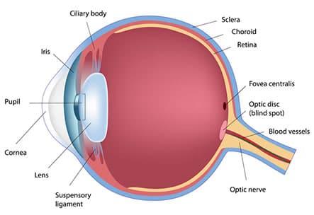
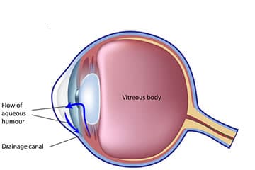
The eye works like a camera: light enters through the cornea, passes through the pupil, and is focused by the lens onto the retina at the back. The retina converts light into signals that travel to the brain along the optic nerve. A clear fluid called aqueous is produced inside the eye to nourish it and maintain its pressure. This fluid drains away through the drainage angle. When drainage is impaired, pressure builds up. Normal eye pressure is between 10 and 21 mmHg.
Symptoms & risk factors
Glaucoma is often called the “sneak thief of sight” because you lose vision without realising it. Early damage affects your peripheral (side) vision, gradually narrowing it to tunnel vision before central sight is affected. Most types progress without any noticeable symptoms until they are advanced — which is why regular screening matters.
Who is most at risk?
- Age over 60
- A family history of glaucoma
- Ethnicity — more common in Caucasians, Asians and Indians
- Short-sightedness or long-sightedness
- A previous eye injury
- High blood pressure or diabetes
- Long-term steroid use
- Migraines
We recommend screening from age 40 if you have risk factors, and from age 45 otherwise.
Types of glaucoma
Primary Open Angle Glaucoma (POAG)
The most common type in New Zealand. A chronic condition where fluid builds up gradually despite an open drainage angle.
Normal Tension Glaucoma (NTG)
Optic nerve damage occurs even though the eye pressure is within the normal range.
Ocular Hypertension (OHT)
Raised eye pressure without any signs of glaucoma yet — monitored closely so treatment can begin if needed.
Acute Angle Closure Glaucoma (ACG)
An emergency. Pressure rises rapidly, causing severe pain, blurred vision and nausea, and needs urgent laser treatment.
Secondary Glaucoma
Glaucoma resulting from another eye condition, such as trauma or inflammation.
How glaucoma is diagnosed
A thorough assessment builds a complete picture of your eye health and may include:
- Vision testing
- Slit-lamp examination of the eye
- Measurement of eye pressure
- Assessment of corneal thickness
- Inspection of the drainage angle
- A dilated examination of the pupil
- Visual field testing
- OCT imaging of the optic nerve
- Stereo-disc photography
Treatment options
Treatment aims to lower the pressure inside the eye to prevent further damage. It’s important to know that treatment prevents further vision loss but cannot restore sight that has already been lost.
Medications (eye drops)
- Prostaglandin analogues
- Beta blockers
- Alpha agonists
- Carbonic anhydrase inhibitors
- Miotics
- Combination drops
Laser procedures
SLT (Selective Laser Trabeculoplasty) improves fluid drainage, with around a 75% success rate and a pressure reduction of roughly 30%. LPI (Laser Peripheral Iridotomy) creates a small opening in the iris for angle closure cases.
Surgical options
- iStent — a minimally invasive device that creates a permanent drainage opening
- Trabeculectomy — creates a bypass hole in the wall of the eye
- ExPRESS device — a modified trabeculectomy using a stainless steel shunt
- Glaucoma tube surgery — inserts a small plastic drainage tube
- Cyclophotocoagulation — reduces fluid production, for more severe cases